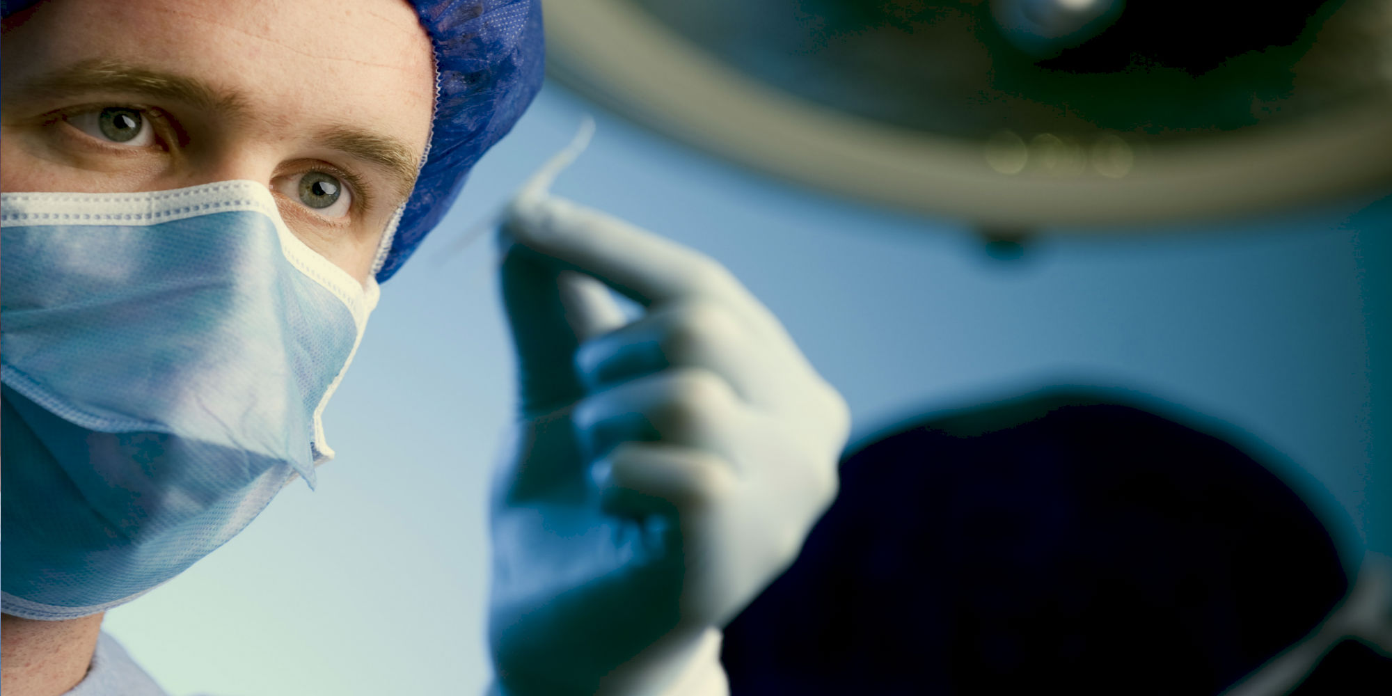
Graduate Research at MDHS
A resource for students and supervisors seeking up to date information about policies and procedures
Working in close partnership with the Royal Children’s Hospital (RCH) and the Murdoch Childrens Research Institute (MCRI), the Department of Paediatrics supports research in all sub-speciality areas of paediatrics (newborn, child, adolescent) medicine, surgery and allied health, across the arenas of clinical, public health and laboratory research - all with the common goal of improving the health outcomes for children today and in the future.
-
Research Themes
Our research is encompassed within several broad research themes. This multidisciplinary approach aims to increase the opportunities for researchers to collaborate on projects that span common themes.
-
Research Groups
An overview of the research interests within the Department and the project work being carried out by our research groups.
-
Honours & Masters Research
We offer research-based courses at Honours and Masters level with a focus on Molecular Biology of Human Development & Disease.
Whether you're just starting out or a seasoned professional, we have something to suit your needs and schedule. With the highest teaching evaluations in the Faculty of Medicine, Dentistry and Health Sciences we are proud to offer our coursework and short course programs to our leaders and future leaders.
-
Degrees
Our degrees, taught by our world-renowned academics are the perfect way to begin in the field of biomedical sciences.In addition to the Child and Adolescent Health component of the Doctor of Medicine we teach a number of post-graduate coursework and research programs.
-
Short Courses and Professional Development
Working with our campus partners at the Melbourne Children’s we manage and deliver a diverse range of non-award short courses, professional development and CME programs to advance healthcare professionals.
-
Scholarships, Bursaries and Prizes
The Faculty of Medicine, Dentistry and Health Sciences offer an extensive range of scholarships and bursaries to undergraduate and postgraduate students.
-
Current Student Resources
Resources, links and helpguides for all enrolled students in the Department.

Clinical Electives
The Department of Paediatrics offers a limited number of places for non-University of Melbourne medical students who wish to undertake clinical electives at the Melbourne Children’s Campus.
-
Teddy Bear Hospital
An innovative vehicle to improve the communication skills of medical professionals and health literacy of children, our Teddy Bear Hospital program is one of the biggest engagement events in the University.
-
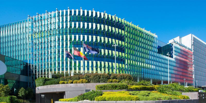
Campus Partners: Melbourne Children’s Campus
The Royal Children’s Hospital, the Murdoch Children’s Research Institute and the University of Melbourne, together make an interwoven, symbiotic relationship delivering high quality clinical services underpinned by research and education.
-
Alumni
Our pride in our graduates and in their lives spent improving the health and well-being of others is matched by our desire to maintain strong connections with all who have passed through our doors. .
ASQ-TRAK
The ASQ-TRAK is a developmental screening tool for observing and monitoring the developmental progress of Australian Aboriginal children at 2 months, 6 months, 12 months, 18 months, 24 months, 36 months and 48 months of age.
Honorary Staff
Our honorary staff play a critical role in the life of the Department of Paediatrics and substantially assist us in working with the communities we serve to improve health and advance health care through our teaching, learning, research, clinical care and advocacy. Applications are welcome from individuals who make a significant and sustained contribution to the Department of Paediatrics in any of the areas of Learning and Teaching; Research; Engagement; or Professional Practice and Leadership.
Welcome
Welcome to the Department of Paediatrics. We are a large and successful department within the Melbourne Medical School of the Faculty of Medicine, Dentistry and Health Sciences with an established worldwide reputation for excellence in child and adolescent health. We are committed to improving clinical care and health outcomes for children and adolescents via our extensive education, research and engagement activities.
Diversity and Inclusion
The Melbourne Medical School is proud to foster a vibrant and inclusive culture delivering initiatives that value and support diversity.

Annual Report 2021-2022
This year's report presents some of the exciting and innovative activities engaged in by our team of students, educators, researchers and leaders. It highlights the achievements of our academic and honorary staff, who continue to generate amazing successes in the face of a global COVID-19 pandemic and its aftermath.